Unit 4 · Botany · 8 teaching hours · 36 min read
Embryology
What the syllabus asks for 7
- Asexual and sexual reproduction in angiosperms
- Pollination
- Fertilization
- Development of male gametophyte
- Development of female gametophyte
- Dicot and monocot embryo
- Endosperm
Embryology is the study of how a flowering plant makes the next generation — how the anther builds pollen, how the ovule builds an egg, how the two meet, and how a single fertilised cell becomes an embryo packed inside a seed.
Everything in this unit happens inside a flower you can pick from a field in Chitwan or Jumla, and almost every stage has a standard labelled diagram that the board asks for by name.
Three things come again and again: (1) a labelled diagram — T.S. of an anther, L.S. of an anatropous ovule, the mature embryo sac, or the Capsella embryo; (2) double fertilization — who fuses with whom, and what ploidy each product has; (3) ploidy counting of every part of a seed.
Learn the sequence microspore mother cell → microspore → pollen grain → male gametes and its female mirror megaspore mother cell → megaspore → embryo sac → egg, and most of the unit falls into place.
4.1 Asexual and sexual reproduction in angiosperms
Angiosperms reproduce in two ways. In asexual reproduction a new plant grows from a part of the parent without gametes and without fertilization, so the offspring is genetically identical to the parent — a clone.
In sexual reproduction two gametes fuse, the chromosome sets of two parents are mixed, and the offspring differs from both.
Asexual reproduction
Vegetative propagation is asexual reproduction by a vegetative organ — root, stem, leaf or bud.
| Organ | Structure | Example |
|---|---|---|
| Underground stem | rhizome | ginger Zingiber officinale, turmeric |
| tuber | potato Solanum tuberosum (eyes = buds) | |
| bulb | onion Allium cepa, garlic | |
| corm | Colocasia, Crocus | |
| Sub-aerial stem | runner | Cynodon dactylon (dubo) |
| sucker | mint Mentha, chrysanthemum | |
| offset | water hyacinth Eichhornia crassipes | |
| Leaf | adventitious buds on the margin | Bryophyllum pinnatum |
| Root | adventitious buds | sweet potato Ipomoea batatas, Dahlia |
Artificial methods used by growers are cutting (sugarcane, rose), layering (jasmine, litchi), grafting and budding (mango, citrus, apple in Mustang and Jumla) and micropropagation by tissue culture.
Apomixis is a special case: a seed is formed without fertilization, so the embryo is a clone of the mother. In nucellar polyembryony in Citrus and mango, extra embryos arise from the nucellus alongside the sexual one.
Apomixis is prized in breeding because it fixes a hybrid's vigour permanently.
| Advantage | Disadvantage | |
|---|---|---|
| Asexual | quick; keeps the parent's exact qualities; works for seedless kinds such as banana | no variation, so the whole crop can fall to one disease; weak strains accumulate |
| Sexual | creates variation, the raw material of breeding and evolution; seeds are easy to store and disperse | slower; needs pollinators; offspring are not identical to the parent |
Sexual reproduction
The flower is the reproductive shoot. The androecium is made of stamens, each a filament bearing an anther; the gynoecium is made of carpels, each a pistil with ovary, style and stigma.
The anther is the microsporangium and produces microspores (pollen grains); the ovule inside the ovary is the megasporangium and produces megaspores. Because the two kinds of spore are different, angiosperms are heterosporous.
The plant body we see is the diploid sporophyte. The pollen grain is the much-reduced male gametophyte and the embryo sac is the female gametophyte; both are haploid, and both are permanently retained inside the sporophyte.
This is the alternation of generations, compressed almost out of sight.
4.2 Pollination
Pollination is the transfer of pollen grains from the anther of a flower to the stigma of a pistil of the same kind of flower.
It is the essential step before fertilization, because the non-motile male gametes of angiosperms can reach the egg only through a pollen tube grown from a grain that has landed on a stigma.
Self-pollination (autogamy) is transfer within the same flower. Geitonogamy is transfer between two flowers of the same plant — genetically still self-pollination, though an agent is needed.
Cross-pollination (xenogamy) is transfer to a flower on a different plant of the same species, and only this brings new genes.
Self-pollination is ensured by homogamy (anther and stigma mature together), by cleistogamy — flowers that never open, as in Commelina benghalensis, Viola and Oxalis — and by bud pollination, as in pea and wheat.
Cross-pollination needs an external agent.
| Agent | Name | Flower characters | Examples |
|---|---|---|---|
| Wind | anemophily | small, dull, no nectar or scent; versatile anthers, huge amounts of light dry pollen; feathery stigma | maize Zea mays, grasses, Pinus, Cannabis |
| Water | hydrophily | pollen and stigma unwettable; pollen often long and thread-like | Vallisneria, Hydrilla, Zostera |
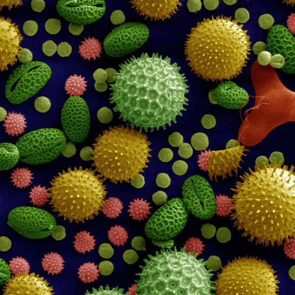
| Insect | entomophily | large, coloured, scented; nectar; sticky spiny pollen; sticky stigma | Salvia, mustard, Ficus (with its wasp) |
| Bird | ornithophily | large, red, tubular, plenty of watery nectar, no scent | Bombax ceiba (simal), Butea monosperma, Erythrina |
| Bat | chiropterophily | large, dull, strong smell, open at night, tough | Kigelia, Adansonia, Anthocephalus |
Because continuous self-pollination narrows the gene pool, plants have evolved contrivances for cross-pollination:
- Dicliny (unisexuality) — separate male and female flowers, either on one plant (monoecious: maize, castor) or on separate plants (dioecious: papaya, Cannabis), which makes selfing impossible in dioecious species.
- Dichogamy — the two sexes ripen at different times: protandry (anthers first) in sunflower Helianthus annuus and Salvia, protogyny (stigma first) in Plantago and Michelia.
- Herkogamy — a physical barrier, such as the pollinia of Calotropis or a stigma held above the anthers.
- Heterostyly — two or three style lengths in the same species, as in Primula and Oxalis, so that pollen is placed at the level of a different plant's stigma.
- Self-incompatibility (self-sterility) — a genetic block that prevents pollen from germinating or the tube from growing on the plant's own stigma, as in tobacco, crucifers and Abutilon.
| Self-pollination | Cross-pollination | |
|---|---|---|
| Agent | not needed | always needed |
| Pollen used | very little | very large amounts wasted |
| Variation | none | plenty |
| Purity of line | maintained | lost |
| Vigour | falls over generations (inbreeding depression) | hybrid vigour |
| Certainty | almost certain | uncertain, depends on the agent |
4.3 Fertilization
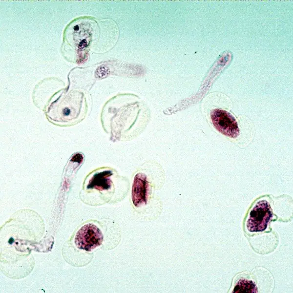
When a compatible pollen grain lands on a receptive stigma it absorbs water and sugars and germinates.
The intine grows out through a germ pore as the pollen tube, which digests its way down through the style, guided chemotropically by substances secreted by the synergids.
The tube nucleus leads at the tip; the generative cell divides (if it has not already) to give two male gametes that follow behind.
The tube reaches the ovule and enters it in one of three ways:
| Route | Name | Example |
|---|---|---|
| Through the micropyle | porogamy | commonest, e.g. most angiosperms |
| Through the chalaza | chalazogamy | Casuarina |
| Through the integument or funicle | mesogamy | Cucurbita, Populus |
The tube then pushes into one synergid, whose filiform apparatus guides it, and bursts, releasing both male gametes into the embryo sac. What follows is unique to angiosperms.
Double fertilization is the fusion of one male gamete with the egg (syngamy, giving a diploid zygote) and of the second male gamete with the two polar nuclei of the central cell (triple fusion, giving a triploid primary endosperm nucleus) in the same embryo sac.
It was discovered by S. G. Nawaschin in 1898 in Lilium and Fritillaria.
Syngamy: male gamete (n) + egg (n) → zygote (2n) → embryo. Triple fusion: male gamete (n) + two polar nuclei (n + n) → primary endosperm nucleus (3n) → endosperm.
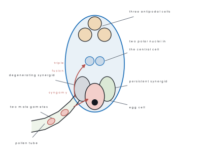
Triple fusion is called triple because three nuclei fuse, not because the product is triploid — though it is.
The significance of double fertilization is that the food store is only laid down after the egg has actually been fertilised, so no plant wastes reserves on an ovule that will never make a seed.
After fertilization the whole flower is rebuilt:
| Before | After |
|---|---|
| Ovule | seed |
| Ovary | fruit |
| Ovary wall | pericarp |
| Integuments | seed coat (testa and tegmen) |
| Zygote | embryo |
| Primary endosperm nucleus | endosperm |
| Nucellus | usually consumed; if it persists, perisperm |
| Micropyle | micropyle of the seed (lets water in at germination) |
| Synergids and antipodals | degenerate |
| Calyx, corolla, stamens, style | usually fall off |
4.4 Development of male gametophyte
The anther
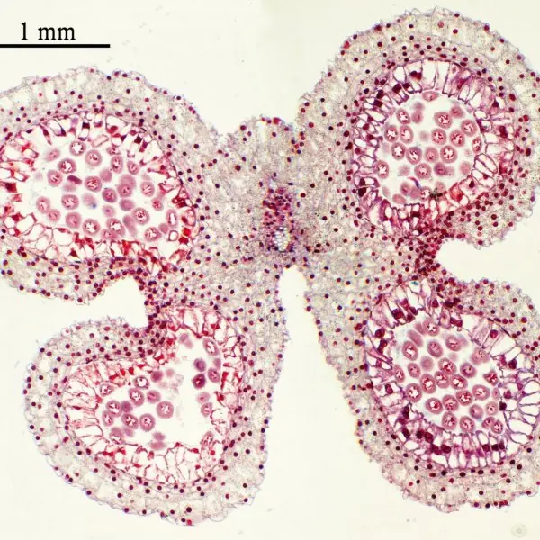
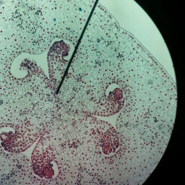
A typical angiosperm anther is dithecous and tetrasporangiate: it has two lobes, and each lobe contains two microsporangia (pollen sacs), so a transverse section is four-lobed and butterfly-shaped.
The two lobes are joined by the connective, through which a vascular bundle runs.
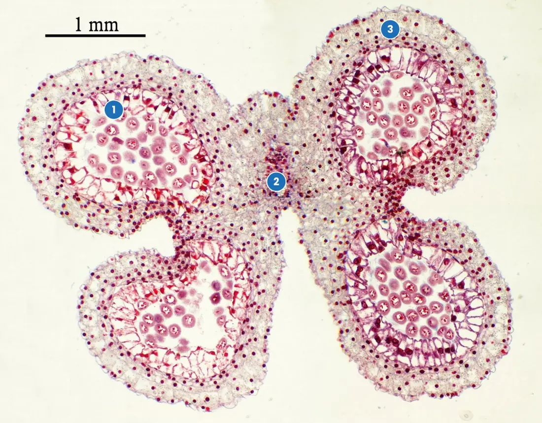
The wall of each microsporangium has four layers, from outside in:
| Layer | Nature | Function |
|---|---|---|
| Epidermis | single, protective | protection |
| Endothecium | single, with fibrous thickenings on the inner and radial walls | hygroscopic; on drying it curls and tears the anther open at the stomium |
| Middle layers | 2–4 thin layers | ephemeral; crushed as pollen grows |
| Tapetum | innermost, cells often bi- or multinucleate, dense cytoplasm | nourishes the developing microspores; supplies sporopollenin for the exine, the pollenkitt and the enzymes and proteins of the pollen wall |
A very common mistake is to give the tapetum the job of protection. Protection is the epidermis; dehiscence is the endothecium; nutrition of the microspores is the tapetum.
If the tapetum fails, the pollen is sterile — which is exactly how male-sterile lines are used in hybrid seed production.
Microsporogenesis and microgametogenesis
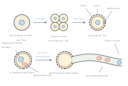
Inside each young sporangium the sporogenous tissue enlarges into microspore mother cells (pollen mother cells, 2n). Each one divides by meiosis to give a tetrad of four haploid microspores. This is microsporogenesis.
The tetrad then separates and each microspore matures into a pollen grain — microgametogenesis.
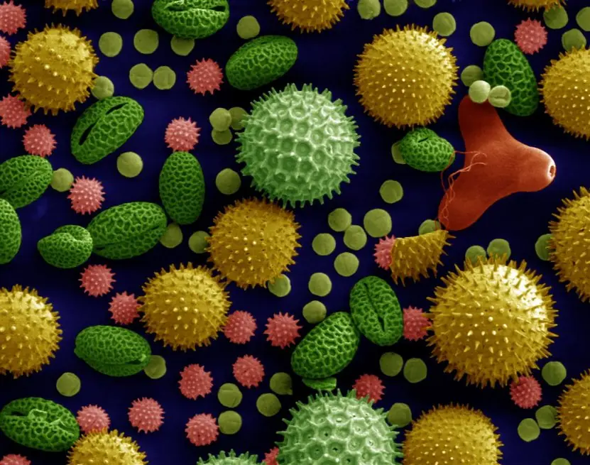
The pollen grain has a two-layered wall: a hard, sculptured outer exine made of sporopollenin, the most resistant organic material known (this is why pollen survives as fossils), interrupted by thin germ pores; and a thin, cellulose-and-pectin intine inside.
The microspore nucleus divides mitotically and very unequally, giving a large vegetative (tube) cell with abundant food and a small, spindle-shaped generative cell floating in its cytoplasm.
In about 60 % of families the pollen is shed at this 2-celled stage and the generative cell divides later inside the pollen tube; in the rest (Poaceae, Asteraceae, Brassicaceae) it divides before shedding, so the grain is shed 3-celled.
Either way, the mature male gametophyte is just three cells — one vegetative cell and two non-motile male gametes.
Problem. An anther of a mustard plant produced 1 200 pollen grains.(a) How many microspore mother cells took part?(b) How many meiotic divisions occurred?(c) How many male gametes can these grains deliver?(d) How many nuclei in all are present in the 1 200 mature (3-celled) grains?
Solution.
(a) Each microspore mother cell gives four microspores, so
(b) One meiosis per mother cell, so 300 meiotic divisions.
(c) Each grain forms two male gametes: 1200 \times 2 = 2400 male gametes.
(d) Each 3-celled grain has one tube nucleus and two male gamete nuclei = 3 nuclei, so 1200 \times 3 = 3600 nuclei.
4.5 Development of female gametophyte
The ovule
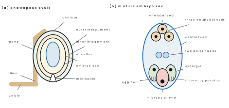
The ovule (megasporangium) is attached to the placenta of the ovary by a stalk, the funicle.
Its parts are the hilum (where funicle meets body), the chalaza (the base of the body, opposite the micropyle), the nucellus (the nutritive tissue), one or two integuments that enclose the nucellus leaving a narrow pore, the micropyle, and, deep inside the nucellus, the embryo sac (female gametophyte).
In an anatropous ovule the funicle is fused along one side as the raphe.
| Type of ovule | Body relative to funicle | Example |
|---|---|---|
| Orthotropous (atropous) | straight, micropyle in line with funicle | Polygonum, Piper, Cycas |
| Anatropous | completely inverted, micropyle beside the hilum — the commonest type, in about 80 % of families | mustard, sunflower, most dicots |
| Campylotropous | body curved, embryo sac slightly curved | Brassica, legumes |
| Amphitropous | body and embryo sac both strongly curved, horseshoe-shaped | Poppy, Alisma |
| Circinotropous | funicle grows right round the ovule | Opuntia |
Megasporogenesis and megagametogenesis
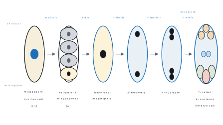
A single hypodermal cell of the nucellus at the micropylar end enlarges as the archesporial cell. In most plants it directly becomes the megaspore mother cell (2n).
This divides by meiosis to give a linear tetrad of four haploid megaspores — that is megasporogenesis. Three of them, usually the three towards the micropyle, degenerate; the chalazal megaspore survives as the functional megaspore.
The functional megaspore now enlarges and its nucleus divides mitotically three times without any wall formation — a free-nuclear stage — giving 2, then 4, then 8 nuclei. Four nuclei lie at each pole.
Walls then form around them: three cells at the micropylar end make the egg apparatus (one egg cell flanked by two synergids whose filiform apparatus guides the pollen tube), three at the chalazal end become the antipodal cells, and the remaining two nuclei — one from each pole — move to the centre as the polar nuclei inside the large central cell.
The result is the Polygonum-type embryo sac: 7 cells and 8 nuclei. It is called monosporic because it develops from a single megaspore, and it occurs in over 80 % of angiosperms.
Problem. A pea pod contains 8 seeds. Assuming no ovule was wasted,(a) how many megaspore mother cells were needed,(b) how many megaspores were formed and how many degenerated,(c) how many mitotic divisions took place in forming the eight embryo sacs, and(d) how many pollen grains were needed at the very least?
Solution.
(a) One seed comes from one ovule, and each ovule has one megaspore mother cell → 8 megaspore mother cells.
(b) Each meiosis gives 4 megaspores, so 8 \times 4 = 32 megaspores were formed and 8 \times 3 = 24 degenerated.
(c) Each functional megaspore undergoes 3 rounds of division: 1 + 2 + 4 = 7 mitotic divisions per embryo sac, so 8 \times 7 = 56 mitotic divisions.
(d) Each pollen grain fertilises one ovule, so at least 8 pollen grains — which would need 8 \div 4 = 2 microspore mother cells.
Problem. Give the ploidy of:(a) nucellus,(b) megaspore mother cell,(c) synergid,(d) antipodal cell,(e) central cell before fertilization, (f) primary endosperm nucleus, (g) endosperm of a maize grain, (h) embryo, (i) seed coat, (j) perisperm of black pepper.
Solution. Work outwards from the rule: everything belonging to the parent sporophyte is 2n, everything in the gametophyte is n, and only the two products of double fertilization differ.
(a) nucellus 2n — sporophyte tissue.(b) megaspore mother cell 2n — it has not yet undergone meiosis.(c) synergid n,(d) antipodal n — both are embryo-sac cells.(e) central cell has two n polar nuclei, so it is 2 × n but has not fused with a gamete. (f) primary endosperm nucleus 3n — n + n + n from triple fusion. (g) endosperm 3n. (h) embryo 2n — from the zygote (n + n). (i) seed coat 2n — it is the integument, mother-plant tissue. (j) perisperm 2n — it is leftover nucellus.
4.6 Dicot and monocot embryo
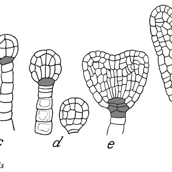
The dicot embryo — Capsella bursa-pastoris
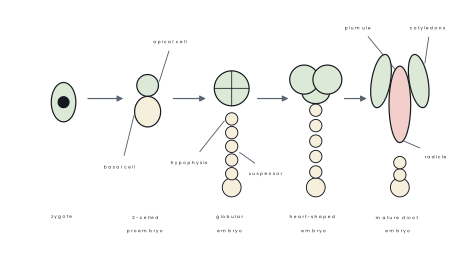
The zygote rests for a while (letting the endosperm get ahead), then divides by a transverse wall into a small apical (terminal) cell towards the chalaza and a large basal cell towards the micropyle.
- The basal cell divides transversely again and again to make a filament of 6–10 cells, the suspensor, which pushes the embryo down into the nourishing endosperm. Its lowest cell swells into a vesicular haustorial cell at the micropyle; its topmost cell, the hypophysis, later forms the tip of the radicle and the root cap.
- The apical cell divides by two vertical walls and one transverse wall into an octant of eight cells. The four epibasal cells make the plumule and the two cotyledons; the four hypobasal cells make the hypocotyl.
- Periclinal divisions then mark out the three primary tissue layers — dermatogen (future epidermis), periblem (cortex) and plerome (stele).
- The embryo passes through a globular, then a heart-shaped stage as the two cotyledon primordia bulge out, then a torpedo stage, and finally curves into the mature embryo.
The monocot embryo
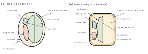
The first division is the same, but the embryo does not form two cotyledons. The mature grass embryo, lying to one side of the endosperm, has:
- a single shield-shaped cotyledon, the scutellum, pressed against the endosperm; its epithelial layer secretes enzymes and absorbs the digested food;
- a coleoptile, a hollow protective sheath over the plumule, which pushes through the soil at germination;
- a coleorhiza, a similar sheath over the radicle;
- an epiblast, a small flap opposite the scutellum, often read as the vestige of the second cotyledon.
| Feature | Dicot embryo | Monocot embryo |
|---|---|---|
| Cotyledons | two, lateral | one, the scutellum, lateral |
| Position | central in the seed | to one side of the endosperm |
| Plumule | terminal, naked | lateral, covered by the coleoptile |
| Radicle | naked | covered by the coleorhiza |
| Epiblast | absent | usually present |
| Food store | usually in the cotyledons (non-endospermic) | in the endosperm (endospermic) |
| Example | Capsella, pea, gram | maize, wheat, rice |
4.7 Endosperm
The endosperm is the triploid (3n) nutritive tissue of an angiosperm seed, formed from the primary endosperm nucleus after triple fusion. It feeds the developing embryo and, in many seeds, the germinating seedling as well.
The endosperm develops in one of three ways.
| Type | How it develops | Examples |
|---|---|---|
| Nuclear | the primary endosperm nucleus divides repeatedly without wall formation, giving a free-nuclear multinucleate mass; walls appear later, from the periphery inwards, or never | commonest type — maize, wheat, rice, Capsella, coconut (the "water" is free-nuclear endosperm and the white kernel is the later cellular part) |
| Cellular | a wall follows every division from the very first, so there is no free-nuclear stage | Datura, Petunia, balsam, Adoxa |
| Helobial | intermediate: the first division is followed by a wall that makes two unequal chambers, then the micropylar chamber goes free-nuclear | order Helobiales — Alisma, Eremurus |
In some seeds the seed coat grows inward in irregular folds, giving a ruminate endosperm, as in betel nut Areca catechu and nutmeg Myristica fragrans.
Seeds are grouped by what happens to the endosperm:
- Endospermic (albuminous) seeds keep the endosperm at maturity, and the cotyledons stay thin: maize, wheat, rice, castor Ricinus communis, coconut. In cereals the outermost endosperm layer is the protein-rich aleurone layer, which on germination secretes α-amylase to digest the starchy endosperm.
- Non-endospermic (exalbuminous) seeds have the endosperm completely absorbed by the cotyledons during seed development, so the fleshy cotyledons themselves are the food store: pea, gram, bean, groundnut, mustard.
If the nucellus also persists in the mature seed as a food store it is called the perisperm, as in black pepper Piper nigrum, beet and cardamom. A perisperm is 2n and is not endosperm at all.
Endosperm is 3n and comes from triple fusion. Cotyledon is 2n and is part of the embryo. Perisperm is 2n and is leftover nucellus. Examiners ask this as a one-mark difference every other year.
The endosperm matters far beyond the plant: the endosperm of wheat, rice and maize is the staple food of most of the world, including the rice grown in the Terai and the maize grown on Nepal's hill terraces.
Wheat flour is almost pure endosperm, and the coconut's liquid endosperm, rich in cytokinins, is used in plant tissue-culture media.

Chapter summary
- Asexual reproduction (rhizome, tuber, bulb, runner, cutting, grafting, apomixis) gives clones — quick and uniform but with no variation; sexual reproduction gives variation.
- Pollination is the transfer of pollen from anther to stigma: autogamy, geitonogamy and xenogamy; agents are wind (anemophily), water (hydrophily), insect (entomophily), bird (ornithophily) and bat (chiropterophily).
- Cross-pollination is enforced by dicliny, dichogamy (protandry and protogyny), herkogamy, heterostyly and self-incompatibility.
- The anther is dithecous and tetrasporangiate; its wall is epidermis, endothecium (dehiscence), middle layers and tapetum (nutrition and sporopollenin).
- Microsporogenesis: microspore mother cell (2n) → meiosis → tetrad of microspores (n). Microgametogenesis: microspore → 2-celled pollen grain (vegetative + generative cell) → two male gametes in the pollen tube.
- Megasporogenesis: megaspore mother cell (2n) → meiosis → linear tetrad; three die, the chalazal one survives. Three free-nuclear mitoses then give the Polygonum-type, monosporic, 7-celled and 8-nucleate embryo sac — egg + 2 synergids, 3 antipodals, and 2 polar nuclei in the central cell.
- Double fertilization (Nawaschin, 1898): syngamy gives the 2n zygote and triple fusion gives the 3n primary endosperm nucleus. Ovule → seed, ovary → fruit, integuments → seed coat, nucellus → perisperm if it persists.
- The dicot embryo (Capsella) passes through 2-celled, octant, globular, heart-shaped and torpedo stages, with a suspensor from the basal cell; the monocot embryo has one cotyledon, the scutellum, plus a coleoptile, a coleorhiza and an epiblast.
- Endosperm development is nuclear (commonest), cellular or helobial. Seeds are endospermic (maize, castor, coconut) or non-endospermic (pea, gram).
Practice questions
Group A — Multiple choice (1 mark each)
- The layer of the anther wall that nourishes the developing microspores is the [1]aepidermisbendotheciumcmiddle layerdtapetum
- A typical angiosperm embryo sac is [1]a7-celled and 8-nucleateb8-celled and 8-nucleatec7-celled and 7-nucleated8-celled and 7-nucleate
- The ploidy of the primary endosperm nucleus is [1]anb2nc3nd4n
- Entry of the pollen tube into the ovule through the chalaza is called [1]aporogamybchalazogamycmesogamydapogamy
- The scutellum of a maize grain is the [1]acoleoptilebendospermcsingle cotyledondperisperm
- Coconut water is [1]afree-nuclear endospermbperispermcnucellusdliquid mesocarp
Answers to Group A
1. (d) — the tapetum supplies food and sporopollenin; the endothecium only helps in dehiscence.
2. (a) — 3 antipodals + 2 synergids + 1 egg + 1 central cell with 2 nuclei = 7 cells, 8 nuclei.
3. (c) — one male gamete (n) fuses with two polar nuclei (n + n).
4. (b) — porogamy is through the micropyle and mesogamy through the integument.
5. (c) — the scutellum is the single, shield-shaped cotyledon of a grass embryo.
6. (a) — it is the free-nuclear stage of nuclear endosperm, so it is 3n.
Group B — Short answer (4 marks each)
- Draw a labelled diagram of a transverse section of a young anther and state the function of each wall layer. [4]
- Describe the development of the male gametophyte from the microspore mother cell to the pollen tube. [4]
- What is double fertilization? Write the two fusions with the ploidy of each product and state its significance. [4]
- An anther contained 250 microspore mother cells. How many pollen grains, male gametes and ovules could it at most fertilise, and how many seeds could result? [4]
- Differentiate between a dicot and a monocot embryo (any four points), and define coleoptile and coleorhiza. [4]
Answers to Group B
1. The section is tetrasporangiate — two lobes, four microsporangia, a central connective with a vascular bundle.
Wall layers from outside in: epidermis — protection; endothecium — fibrous thickenings that dry and curl, tearing the anther open at the stomium; middle layers — 2–4 short-lived layers, crushed as pollen grows; tapetum — innermost, multinucleate, supplies nutrition, sporopollenin, pollenkitt and enzymes to the microspores.
2. Sporogenous tissue → microspore mother cells (2n) → meiosis → tetrad of four haploid microspores (microsporogenesis). Each microspore builds a two-layered wall — sporopollenin exine with germ pores, and cellulose intine.
Its nucleus then divides unequally by mitosis into a large vegetative (tube) cell and a small generative cell; the grain is usually shed at this 2-celled stage.
On the stigma the grain germinates, the intine grows out through a germ pore as the pollen tube, and the generative cell divides into two male gametes, which travel down the tube behind the tube nucleus.
3. Double fertilization is the occurrence of two fusions in the same embryo sac. Syngamy: one male gamete (n) + egg (n) → zygote (2n) → embryo.
Triple fusion: the second male gamete (n) + two polar nuclei (n + n) → primary endosperm nucleus (3n) → endosperm. It was discovered by Nawaschin (1898).
Significance the food-storing endosperm is laid down only after the egg is fertilised, so no reserves are wasted on an ovule that will not become a seed; the triploid endosperm is also vigorous and gives the young seedling a strong start. It is found only in angiosperms.
4. Each microspore mother cell gives 4 pollen grains:
Each grain forms two male gametes: 1000 \times 2 = 2000 male gametes. But only one grain fertilises one ovule (the second gamete goes to the polar nuclei), so these grains could fertilise at most 1000 ovules and give at most 1000 seeds.
5.
| Feature | Dicot embryo | Monocot embryo |
|---|---|---|
| Number of cotyledons | two | one (scutellum) |
| Position of cotyledons | terminal, on either side of the axis | lateral, to one side |
| Plumule | naked | enclosed in a coleoptile |
| Radicle | naked | enclosed in a coleorhiza |
| Epiblast | absent | usually present |
The coleoptile is the hollow protective sheath over the plumule of a grass embryo, and the coleorhiza is the protective sheath over its radicle.
Group C — Long answer (8 marks each)
- Describe the structure of a typical anatropous ovule with a labelled diagram, and explain the development of the female gametophyte from the megaspore mother cell to the mature embryo sac. [8]
- What is pollination? Describe the different agents of cross-pollination with the flower characters and examples of each, and explain any four contrivances that plants use to ensure cross-pollination. [8]
- Describe the development of a dicot embryo in Capsella bursa-pastoris with labelled diagrams, and compare it with a monocot embryo. What is endosperm, and how do the three types of endosperm development differ? [8]
Answers to Group C
1. Outline. (i) The ovule is attached to the placenta by the funicle; the point of attachment to the body is the hilum, and in an anatropous ovule the funicle is fused along one side as the raphe, so that the body is completely inverted and the micropyle lies right beside the hilum. (ii) The body consists of the nucellus, surrounded by one or two integuments which leave the micropyle open at one end, and the chalaza at the opposite end. The embryo sac lies within the nucellus. (iii) A hypodermal archesporial cell at the micropylar end becomes the megaspore mother cell (2n). (iv) Megasporogenesis: meiosis gives a linear tetrad of four haploid megaspores; the three nearer the micropyle degenerate and the chalazal megaspore is functional. (v) Megagametogenesis: the functional megaspore enlarges and its nucleus divides mitotically three times without wall formation, giving 2, then 4, then 8 nuclei, four at each pole. (vi) Walls then form: three cells at the micropylar end make the egg apparatus (egg + two synergids with a filiform apparatus), three at the chalazal end become antipodal cells, and one nucleus from each pole moves to the centre as the two polar nuclei in the large central cell. (vii) The mature sac is therefore 7-celled and 8-nucleate, monosporic, and is called the Polygonum type. A labelled diagram of the ovule and of the embryo sac carries most of the marks.
2. Outline. (i) Definition — pollination is the transfer of pollen from the anther to the stigma; self (autogamy), geitonogamy and cross (xenogamy).
(ii) Anemophily — wind: small, dull, scentless flowers, versatile anthers, enormous amounts of light dry pollen, feathery stigmas — maize, grasses.
(iii) Hydrophily — water: unwettable pollen, often thread-like — Vallisneria, Hydrilla, Zostera.
(iv) Entomophily — insects: large, brightly coloured, scented flowers with nectar, sticky spiny pollen and sticky stigma — Salvia, mustard, Ficus.
(v) Ornithophily — birds: large red tubular flowers with copious watery nectar and no scent — Bombax ceiba, Butea monosperma. (vi) Chiropterophily — bats: large, dull, strong-smelling, night-opening flowers — Kigelia, Adansonia.
(vii) Contrivances (any four): dicliny (unisexual flowers, monoecious maize or dioecious papaya); dichogamy (protandry in sunflower, protogyny in Plantago); herkogamy (a physical barrier, as in Calotropis); heterostyly (different style lengths in Primula); self-incompatibility (a genetic block, as in tobacco).
3. Outline. (i) The zygote divides transversely into a small apical cell and a large basal cell.
(ii) The basal cell forms the suspensor, whose lowest cell becomes the vesicular haustorium and whose top cell, the hypophysis, gives the radicle tip.
(iii) The apical cell divides by two vertical and one transverse wall into an octant; the four epibasal cells give the plumule and cotyledons, the four hypobasal cells give the hypocotyl.
(iv) Periclinal divisions give dermatogen, periblem and plerome. (v) The embryo passes through globular → heart-shaped → torpedo → mature stages, ending with two cotyledons, a plumule and a radicle. (vi)
Comparison a monocot embryo has one lateral cotyledon, the scutellum, with a coleoptile over the plumule, a coleorhiza over the radicle and an epiblast; its food is in the endosperm, while a dicot usually stores food in the cotyledons.
(vii) Endosperm is the 3n nutritive tissue formed from the primary endosperm nucleus.
(viii) Nuclear type — repeated free-nuclear divisions with walls laid down later or never (maize, coconut); cellular type — a wall after every division from the first (Datura, Petunia); helobial type — one wall makes two unequal chambers and the micropylar chamber then goes free-nuclear (Alisma, Helobiales).
Group D — Model answers for long questions (10 marks each, for deeper revision)
- Describe the development of the male gametophyte (microsporogenesis and microgametogenesis) from the structure of the anther. [10]
- Describe the development of the female gametophyte (embryo sac) in a typical angiosperm and draw a labelled mature embryo sac. [10]
- What is double fertilization? Explain the process, the formation of endosperm and embryo, and give eight points of its significance. [10]
Model answer D1: development of the male gametophyte (10 marks)
Introduction. The male gametophyte of an angiosperm is the pollen grain. It develops inside the anther from diploid cells by meiosis and mitosis, and it is reduced to only two or three cells.
Structure of the anther. A typical anther is bilobed and tetrasporangiate: it has four pollen sacs (microsporangia).
The wall has four layers from outside inwards: the epidermis (protection), the endothecium (fibrous thickenings that help the anther to dehisce), the middle layers (which degenerate) and the tapetum, a nutritive layer that supplies food, enzymes and sporopollenin precursors to the developing pollen. Inside is the sporogenous tissue.
Stage 1: microsporogenesis (formation of microspores).
- Each cell of the sporogenous tissue becomes a microspore mother cell (pollen mother cell, 2n).
- Each mother cell divides by meiosis, giving a tetrad of four haploid microspores (n).
- The four microspores separate and are released as individual cells. A single anther of a lily produces thousands of pollen grains in this way.
Stage 2: microgametogenesis (formation of the male gametophyte).
- The microspore enlarges and develops a two-layered wall: an outer exine made of sporopollenin, one of the most resistant organic materials known, and an inner intine of cellulose and pectin. The exine has thin places called germ pores.
- The nucleus divides unequally by mitosis to give a large vegetative cell and a small generative cell.
- At this stage the pollen is shed in about 60% of species (the two-celled stage). In the rest, the generative cell divides again to produce two male gametes before the pollen leaves the anther (the three-celled stage, as in maize and wheat).
- If the pollen lands on a compatible stigma, the vegetative cell forms the pollen tube and the two male gametes pass into it.
| Feature | Microsporogenesis | Microgametogenesis |
|---|---|---|
| Starting cell | Pollen mother cell (2n) | Microspore (n) |
| Division | Meiosis | Mitosis |
| Product | Tetrad of 4 microspores | Pollen grain with vegetative and generative cells |
| Chromosome number | 2n → n | n → n |
Importance of the tapetum and the exine. Without the tapetum, pollen is sterile; a failure of its development is used to make male-sterile lines for hybrid seed. The exine protects the grain from drying, and it preserves pollen as fossils.
Conclusion. One diploid pollen mother cell therefore gives four pollen grains, each a two- or three-celled haploid male gametophyte ready for pollination.
Model answer D2: development of the female gametophyte (10 marks)
Introduction. The female gametophyte is the embryo sac, formed inside the ovule. In about 70% of flowering plants it is of the monosporic, Polygonum type, which develops from one megaspore.
Structure of the ovule. The ovule is joined to the placenta by a stalk (funicle). It has a mass of tissue called the nucellus surrounded by one or two integuments that leave a small opening, the micropyle.
The base where the integuments and nucellus meet is the chalaza. A single megaspore mother cell (2n) lies in the nucellus below the micropylar end.
Stage 1: megasporogenesis. The megaspore mother cell divides by meiosis into a linear tetrad of four haploid megaspores. In the Polygonum type, the three towards the micropyle degenerate, and the one near the chalaza becomes the functional megaspore.
Stage 2: megagametogenesis.
- The functional megaspore enlarges, and its nucleus divides by mitosis to form two nuclei, which move to opposite (micropylar and chalazal) poles.
- Each of the two nuclei divides twice by mitosis, and a total of four nuclei at each pole (8 nuclei) are formed. This is a free nuclear division with no walls.
- Three nuclei at each pole become surrounded by cell walls, and one nucleus from each pole, the polar nuclei, moves to the centre.
- The result is a 7-celled, 8-nucleate embryo sac.
| Part | Position | Number of cells and nuclei | Function |
|---|---|---|---|
| Egg apparatus: egg cell | Micropylar end | 1 cell | Fuses with male gamete to form zygote |
| Synergids | Beside the egg | 2 cells | Secrete chemicals that guide the pollen tube (filiform apparatus) |
| Central cell | Centre | 1 cell, 2 polar nuclei | Fuses with the second male gamete to form the endosperm |
| Antipodal cells | Chalazal end | 3 cells | Nourish the embryo sac; degenerate after fertilization |
Counting. 3 (egg apparatus) + 3 (antipodals) + 1 (central cell) = 7 cells; 3 + 3 + 2 = 8 nuclei. All are haploid, with the central cell having two nuclei and therefore becoming triploid after fusion with a male gamete.
Diagram guide. Draw an oval sac with the three-celled egg apparatus at the micropylar end (egg in the middle, a synergid on either side), a large central cell with two polar nuclei, and three small antipodal cells at the chalazal end.
Conclusion. One diploid megaspore mother cell gives one functional megaspore and, after three mitoses, a mature seven-celled embryo sac, ready for double fertilization.
Model answer D3: double fertilization and its significance (10 marks)
Introduction. Double fertilization is the event in which one male gamete fuses with the egg (syngamy) and the other with the two polar nuclei (triple fusion) in the same embryo sac.
It is found only in angiosperms and was discovered by Nawaschin (1898) in Lilium and Fritillaria.
The process.
- A pollen grain lands on the stigma and absorbs water, and the vegetative cell produces a pollen tube through a germ pore.
- The tube grows down the style, guided by chemicals from the synergids, and enters the ovule most commonly through the micropyle (porogamy).
- It enters one synergid and releases the two male gametes into the embryo sac.
- Syngamy: one male gamete (n) fuses with the egg (n) to give the zygote (2n), which develops into the embryo.
- Triple fusion: the other male gamete (n) fuses with the two polar nuclei (n + n) to form the primary endosperm nucleus (3n), which develops into the endosperm.
- After fertilization the synergids and antipodals degenerate, the ovule becomes the seed, the integuments form the seed coat and the ovary becomes the fruit.
Significance (8 points).
- Saves food. The endosperm begins to develop only after fertilization, so the plant does not waste food on unfertilized ovules.
- Provides food for the embryo. The triploid endosperm stores starch, protein and oil that nourish the seedling, as in the rice and wheat grains that feed Nepal.
- Restores the diploid number. Syngamy brings the chromosome number back from n to 2n.
- Gives variation. Two gametes from different parents mix their genes and make new combinations.
- Triploid tissue is vigorous. The extra set of chromosomes in the endosperm gives rapid growth and heterosis-like vigour.
- Ensures coordination. The embryo and its food supply start together, so the seed is ready in a single step.
- Starts fruit development. After fertilization hormones from the seed make the ovary grow into the fruit.
- Basis for breeding. Understanding it allows controlled crosses, seed-set studies and embryo rescue in hybrids.
Conclusion. Double fertilization joins the formation of the embryo with that of its food, and this efficient reproductive system helps to explain the success of flowering plants.
Practise more: Biology question bank